
Grade 10 Life Sciences — June 2025
NATIONAL SENIOR CERTIFICATE
GRADE 10
LIFE SCIENCES
JUNE COMMON TEST — 2025
MARKS: 150
TIME: 2½ Hours
INSTRUCTIONS AND INFORMATION
Read the following instructions carefully before answering the questions.
- Answer ALL the questions.
- Write ALL the answers in the ANSWER BOOK.
- Start the answers to each question at the top of a NEW page.
- Number the answers correctly according to the numbering system used in this question paper.
- Present your answers according to the instructions of each question.
- Do ALL drawings in pencil and label them in blue or black ink.
- Draw diagrams, tables or flow charts only when asked to do so.
- The diagrams in this question paper are NOT necessarily drawn to scale.
- Do NOT use graph paper.
- You may use a non-programmable calculator, protractor and a compass.
- Write neatly and legibly.
SECTION A
QUESTION 1
Various options are provided as possible answers to the following questions. Choose the correct answer and write only the letter (A – D) next to the question number (1.1.1 to 1.1.10) in your ANSWER BOOK, for example 1.1.1 D.
The blood vessel that carries blood to the kidneys is the …
SHOW ANSWER
Answer
D — renal artery.
The renal artery carries blood to the kidneys.
The renal artery carries blood from the heart towards the kidneys for filtration and other kidney functions.
The first scientist to see living cells using microscope is …
SHOW ANSWER
Answer
C — Antonie Van Leeuwenhoek.
Antonie Van Leeuwenhoek is associated with the early observation of living microscopic cells using microscopes.
The chemical used to test for glucose is …
SHOW ANSWER
Answer
B — Benedict's solution.
Benedict's solution is used to test for glucose and other reducing sugars.
The following elements are found in the organic compounds:
Which ONE of the following is the CORRECT combination of elements found in carbohydrates?
SHOW ANSWER
Answer
D — (i), (iii), (iv) only.
Carbohydrates contain carbon, hydrogen and oxygen, represented by statements (iv), (i) and (iii).
Which ONE of the following parts form the axial skeleton?
SHOW ANSWER
Answer
D — Skull, vertebral column, ribs and sternum.
The axial skeleton forms the central framework of the body and includes the skull, vertebral column, ribs and sternum.
The type/s of skeleton/s found in snail is/ are …
SHOW ANSWER
Answer
B/C — exoskeleton and endoskeleton.
The answer shown preserves the source's stated alternatives for the type or types of skeleton in a snail.
The diagram below shows a phase in mitosis.
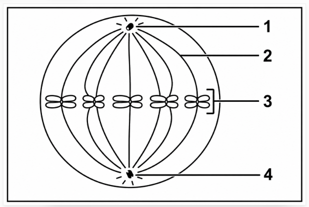
| 1 | 2 | 3 | 4 | |
|---|---|---|---|---|
| A | Nuclear membrane | Spindle fibres | Chromosome | Centrosome |
| B | Cell membrane | Chromosome | Centrosome | Spindle fibres |
| C | Nuclear membrane | Centrosome | Chromosome | Spindle fibres |
| D | Nuclear membrane | Spindle fibre | Centrosome | Chromosome |
SHOW ANSWER
Answer
No answer is provided for this item in the supplied source. No answer.
No answer is provided for this item in the supplied source.
The instrument used to measure the rate of transpiration is called …
SHOW ANSWER
Answer
B — potometer.
A potometer is used to estimate the rate of water uptake associated with transpiration.
Which of the following consist of sclerenchyma cap, phloem, cambium, and xylem?
SHOW ANSWER
Answer
C — Vascular bundle.
A vascular bundle contains conducting tissues including xylem and phloem, together with associated tissues.
Which of the following parts form part of phloem tissue?
SHOW ANSWER
Answer
C — Sieve tubes and companion cells.
Sieve tubes and companion cells are the characteristic conducting components identified as part of phloem tissue.
(10 × 2) (20)
Give the correct biological term for each of the following descriptions. Write only the term next to the question number (1.2.1 to 1.2.9) in your ANSWER BOOK.
SHOW ANSWER
Answer
Polysaccharides
Polysaccharides are large carbohydrate molecules formed when many monosaccharide units are joined together.
SHOW ANSWER
Answer
Goblet cells
Goblet cells are specialised cells that produce mucus, helping to protect and lubricate the epithelial surface.
SHOW ANSWER
Answer
Amino acids
Proteins are built from smaller units called amino acids, which are joined together to form protein molecules.
SHOW ANSWER
Answer
Chromoplast
Chromoplasts contain pigments that give plant parts red, yellow and orange colours.
SHOW ANSWER
Answer
Cranium
The cranium forms the part of the skull that surrounds and protects the brain.
SHOW ANSWER
Answer
Atrium / atria
The atria are the upper chambers of the heart and receive blood returning to the heart.
SHOW ANSWER
Answer
Fibrous / connective tissue
The dense collagen fibres give fibrous connective tissue strength and support.
SHOW ANSWER
Answer
Phloem
Phloem transports manufactured food from the leaves to other parts of the plant.
SHOW ANSWER
Answer
Stem
In the stem, the vascular bundles are arranged in a ring, which is a characteristic used to identify this plant organ.
(9 × 1) (9)
Indicate whether each of the statements in COLUMN I applies to A ONLY, B ONLY, BOTH A AND B or NONE of the items in COLUMN II. Write A only, B only, both A and B, or none, next to the question number (1.3.1 to 1.3.4) in the ANSWER BOOK.
| Column I | Column II |
|---|---|
| 1.3.1 Tissue that joins bone to bone | A: Ligaments B: Tendons |
| 1.3.2 Provides mechanical support to the plant | A: Sclerenchyma B: Collenchyma |
| 1.3.3 Contains digestive enzymes | A: Lysosomes B: Golgi body |
| 1.3.4 Contains bone cells in hard matrix | A: Cartilage B: Ligaments |
SHOW ANSWER
Answer
A only
Ligaments join bone to bone, whereas tendons connect muscle to bone.
SHOW ANSWER
Answer
Both A and B
Both sclerenchyma and collenchyma contribute mechanical support to plant tissues.
SHOW ANSWER
Answer
A only
Lysosomes contain digestive enzymes that break down materials inside cells.
SHOW ANSWER
Answer
None
Neither cartilage nor ligaments contains bone cells embedded in a hard bone matrix.
(4 × 2) (8)
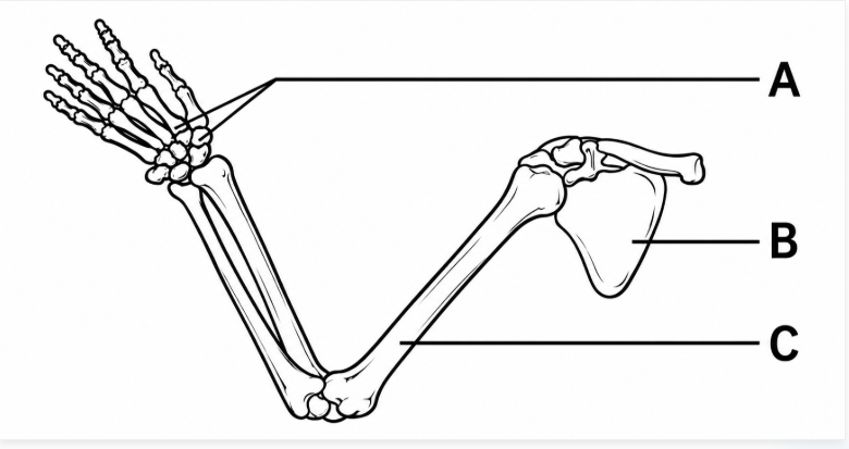
The diagram below shows part of a human skeleton.
SHOW ANSWER
Answer
B — Scapula
The scapula is the shoulder blade and forms part of the pectoral region of the appendicular skeleton.
SHOW ANSWER
Answer
C — Humerus
The humerus is the long bone of the upper arm and connects the shoulder region to the elbow.
SHOW ANSWER
Answer
A — metacarpals
The metacarpals form the framework of the hand between the wrist bones and the finger bones.
SHOW ANSWER
Answer
Appendicular skeleton
The appendicular skeleton includes the limb bones and the girdles that attach the limbs to the axial skeleton.
SHOW ANSWER
Answer
No answer is provided for this item.
No answer is provided for this item in the supplied source.
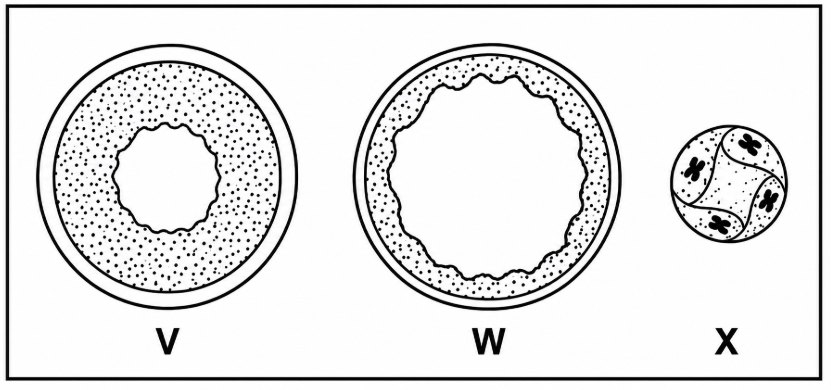
The diagrams below show cross sections through three types of blood vessels.
SHOW ANSWER
Answer
W — Vein
A vein carries blood towards the heart and has structural features suited to return blood at relatively low pressure.
SHOW ANSWER
Answer
X — Capillary
Capillaries are tiny vessels with very thin walls that allow exchange between blood and surrounding tissues.
SHOW ANSWER
Answer
V — Artery
An artery has thick muscular and elastic walls because it carries blood away from the heart under relatively high pressure.
SHOW ANSWER
Answer
W — Vein
Veins carry blood towards the heart.
SECTION B
QUESTION 2
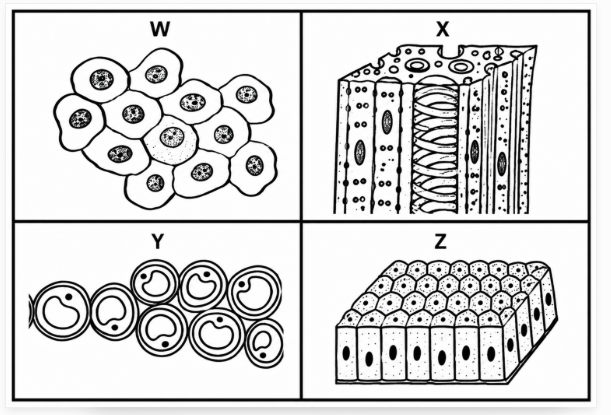
The diagrams below show animal and plant tissues.
SHOW ANSWER
Answer
Y — parenchyma
Parenchyma is a plant tissue whose relatively unspecialised cells can perform storage and other basic tissue functions.
SHOW ANSWER
Answer
Z — columnar epithelium
Columnar epithelium consists of elongated cells arranged in a layer and is found in locations such as the stomach and intestines.
SHOW ANSWER
Answer
Thin / flat / single layer of cells lining blood vessels / lungs.
A thin single layer gives a short diffusion distance, which suits tissue lining areas where substances need to move across the surface.
SHOW ANSWER
Answer
Long / elongated / single layer of cells lining the stomach / intestines / parts of respiratory tracts.
The elongated cells form a single epithelial layer in locations where this tissue carries out its specialised lining function.
SHOW ANSWER
Answer
- Tissue Y acts as a packing tissue.
- It stores food, water and other substances.
- It allows movement of water and gases through the intercellular spaces.
Parenchyma can fill spaces within plant organs, store substances and allow movement through the spaces between its cells.
SHOW ANSWER
Answer
- The cells are elongated and the cross walls disappear to form continuous tubes for the transport of water.
- Xylem tissue does not contain cytoplasm / contains no living tissue so that the water is not blocked by the cytoplasm.
- The cells are strengthened with lignin, protecting them from collapsing under strong suction pressure.
These structural features allow xylem to form strong, continuous pathways for water movement while reducing obstruction and preventing the vessels from collapsing.
Grade 10 learners conducted an investigation to illustrate the movement of water across a visking tubing. (Visking tubing allows water to move in and out like the cell membrane of the cell).
The learners:
- Filled the visking tubing with strong sucrose solution.
- Placed the filled visking tubing in a beaker with tap water.
- Recorded the level of water in the beaker at the beginning of investigation.
- The set up was left in the laboratory for 2 hours.
- After 2 hours there was a change in the level of water in the beaker and the solution in the visking tubing.
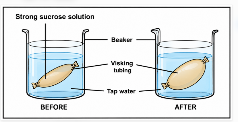
(a) ONE reason for recording the water levels in the beaker at the beginning of the investigation. (2)
(b) TWO changes that are shown in the level of the strong sucrose solution after 2 hours. (4)
(c) Why the sugar molecules did not move from the visking tubing to the beaker. (2)
SHOW ANSWER
Answer
Osmosis.
Osmosis is the movement of water across a selectively permeable membrane, as demonstrated by the change in the visking tubing setup.
SHOW ANSWER
Answer
| Passive transport | Active transport |
|---|---|
| Does not require energy / carrier molecule | Requires energy / carrier molecule |
| Movement along concentration gradient | Movement against concentration gradient |
Passive transport moves substances without the stated energy requirement, while active transport requires energy and can move substances against the concentration gradient.
SHOW ANSWER
Answer
To serve as a baseline / reference point where the results will be compared after two hours, so that the change in the water level can be determined.
A starting measurement provides a reference against which the later water level can be compared to determine the change.
SHOW ANSWER
Answer
There is an increase in the amount of solution in the visking tubing. Since the water has moved from the beaker into the visking tubing through the selectively permeable membrane, the level of solution in the visking tubing increases.
Water moves into the visking tubing through the selectively permeable membrane, increasing the amount and level of solution inside it.
SHOW ANSWER
Answer
The sugar molecules are too large to pass through the selectively permeable membrane of the visking tubing.
The selectively permeable membrane allows water to pass but prevents the larger sugar molecules from passing through.
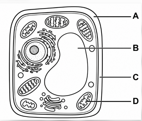
The diagram below shows a type of a cell.
SHOW ANSWER
Answer
Plant cell
The diagram represents a plant cell, which has characteristic structures such as a cell wall, chloroplasts and a large vacuole.
SHOW ANSWER
Answer
B — Vacuole
The vacuole is a large fluid-filled compartment in a typical plant cell.
SHOW ANSWER
Answer
C — Cell wall
The cell wall forms a rigid outer boundary that supports and protects the plant cell.
SHOW ANSWER
Answer
- Cell wall
- Chloroplasts
- Regular shape
- Large vacuole
These visible features distinguish a typical plant cell from an animal cell. The complete set of valid points is shown so the learner can study the available evidence.
SHOW ANSWER
Answer
- Provides support to the plant cell.
- Serves as a storage organ / stores water / salts.
The vacuole stores substances and helps maintain the internal pressure that supports the plant cell.
SHOW ANSWER
Answer
During cellular respiration part D / mitochondrion produces / stores energy, which will be used by the cell for its functions / metabolic processes.
The mitochondrion is involved in cellular respiration, providing energy for the cell's functions and metabolic processes.
SHOW ANSWER
Answer
There will be no movement of substances in and out of the cell; useful substances will become a shortage inside the cell; waste material will also accumulate inside the cell, resulting in the death of the cell / cell becoming dysfunctional.
If the membrane becomes impermeable, substances cannot be exchanged normally. Useful substances become limited and wastes accumulate, so normal cell function is eventually disrupted.
Grade 10 learners conducted an investigation to determine the effect of temperature on the action of pepsin (protein-digestive enzyme).
The procedure was as follow:
- 20 ml of stomach fluid and 10 grams of protein were placed in each of five test tubes.
- The tubes were then kept at different temperatures.
- After 24 hours, the contents of each tube were tested to determine the amount of protein that had been digested.
| Temperature (°C) | Amount of protein (g) |
|---|---|
| 5 | 0.5 |
| 10 | 1.0 |
| 20 | 4.0 |
| 37 | 9.5 |
| 85 | 0.0 |
SHOW ANSWER
Answer
Amount of protein digested.
The dependent variable is the result being measured in response to the temperature conditions.
SHOW ANSWER
Answer
37°C.
The greatest amount of protein was digested at 37°C, so this temperature is the optimum shown by the investigation.
SHOW ANSWER
Answer
- Increase the sample size / amount of stomach fluid and proteins.
- Repeat investigation / several times.
Increasing the amount of material and repeating the investigation provide more observations and help make the result more reliable.
SHOW ANSWER
Answer
Percentage increase = ((9.5 − 4.0) ÷ 4.0) × 100 = 137.5%.
The percentage increase compares the increase from the 20°C value with the original 20°C value, then expresses that change as a percentage.
SHOW ANSWER
Answer
The starch would not be digested.
The stated investigation concerns pepsin, a protein-digestive enzyme, so the outcome given for starch is that it would not be digested.
SHOW ANSWER
Answer
No answer is provided for this item.
No answer is provided for this item in the supplied source.
SHOW ANSWER
Answer
No answer is provided for this item.
No answer is provided for this item in the supplied source.
QUESTION 3
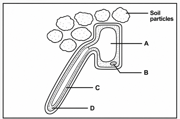
The diagram below shows a root hair.
SHOW ANSWER
Answer
A — Vacuole.
The vacuole is the labelled internal space in the root hair cell and contains cell sap.
SHOW ANSWER
Answer
B — Controls all the activities of the cell / contains DNA which controls hereditary characteristics.
The nucleus contains genetic material and controls the activities of the cell.
SHOW ANSWER
Answer
Root hair region.
Root hairs occur in the specialised root hair region, where they provide the surface used for absorption.
SHOW ANSWER
Answer
- Root pressure
- Transpiration pull
- Capillarity
These three forces contribute to the upward movement of water through the plant from the roots towards the leaves.
SHOW ANSWER
Answer
Part C / cell wall of a root hair is fully permeable and allows both salt and water to pass through, whereas part D / cell membrane of a root hair is semi-permeable and does not allow molecules of salt to pass through.
The cell wall allows substances to pass through freely, while the selectively permeable cell membrane controls which substances can cross.
SHOW ANSWER
Answer
- It has finger-like projections, which increase surface area for absorption of water.
- It has a thin cell wall, allowing it to absorb water from the soil.
- It has low water potential in the cell sap, increasing the water absorption potential.
The long projection increases surface area, the thin wall shortens the pathway for water, and the low water potential supports water absorption from the soil.
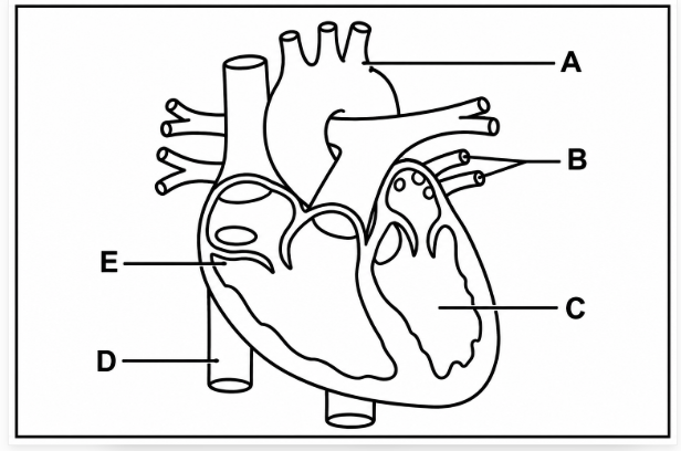
The diagram below shows the human heart.
SHOW ANSWER
Answer
D.
Part D is the vessel carrying deoxygenated blood from the lower parts of the body back towards the heart.
SHOW ANSWER
Answer
Oxygenated blood returns from the lungs → enters the heart through the pulmonary veins → passes into the left atrium → passes the bicuspid/mitral valve → enters the left ventricle → which pumps the blood to part A / aorta.
The blood returns oxygenated from the lungs, enters the left side of the heart, passes through the mitral valve into the left ventricle, and is then pumped into the aorta.
SHOW ANSWER
Answer
Part C / left ventricle is thicker and more muscular / larger, while part E / right ventricle is thinner and less muscular.
The left ventricle has a thicker muscular wall because it must pump blood out to the body, whereas the right ventricle pumps blood to the lungs.
SHOW ANSWER
Answer
The heart is starved of oxygen; since the blood with oxygen cannot enter, this leads to serious heart damage such as heart attack / death of heart muscle.
A blockage reduces the oxygen supply to heart muscle. Prolonged oxygen shortage can damage the muscle and may result in a heart attack or death of heart muscle.
Grade 10 learners were given the set up to use as shown below.
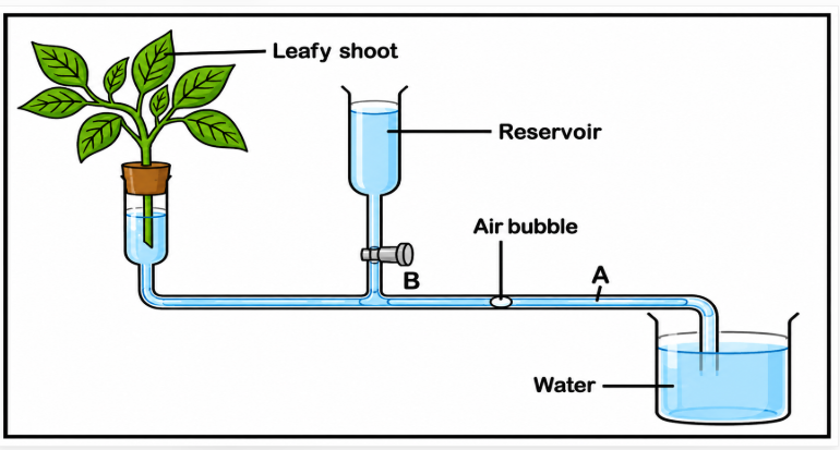
Learners had to:
- Give the leafy shoot some time to adapt to a new environmental condition before taking readings.
- Introduce an air bubble into the capillary tubing and time how long it takes to move from point A to B.
- To repeat the procedure several times.
- Use the reservoir with the tap to move the air bubble back to point A before it was timed again.
- The table below shows the average results learners obtained after repeating the procedure three times.
| Environmental conditions | Average time taken for air bubble to move 5 cm (seconds) |
|---|---|
| Laboratory | 155 |
| Bright light | 125 |
| Wind / fan | 135 |
| Plastic bag | 160 |
SHOW ANSWER
Answer
To measure the rate of transpiration under different environmental conditions.
The investigation compares transpiration under different environmental conditions by measuring how quickly the air bubble moves.
SHOW ANSWER
Answer
Plastic bag.
The plastic-bag condition had the longest average time for the air bubble to move 5 cm, corresponding to the lowest transpiration rate in the investigation.
SHOW ANSWER
Answer
No answer is provided for this item.
No answer is provided for this item in the supplied source.
SHOW ANSWER
Answer
Same leafy twig was used.
Keeping the same leafy twig constant helps ensure that the environmental condition is the factor being compared.
SHOW ANSWER
Answer
Leafy twig shoot should be cut at oblique angle to increase surface area for water absorption.
Cutting the shoot obliquely increases the cut surface area available for water absorption.
SHOW ANSWER
Answer
The stomata would be clogged / blocked, causing the leaf to retain water and stopping transpiration.
Blocking the stomata reduces the route through which water vapour leaves the leaf, so transpiration is reduced or stopped.
SHOW ANSWER
Answer
Model answer graph:
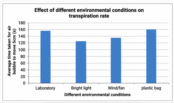
Graph requirements: use a bar graph; include a caption containing both variables; label the X-axis and Y-axis correctly with units; use equal-width bars with equal spaces; and plot all four values correctly.
The model graph shows the four environmental conditions and their average times, with the axes, units and bars presented so the data can be compared.
Read the extract below.
Leukemia and Bone Marrow Transplant
Leukemia is type of cancer which affects blood cells. This disease (as well as many others) is caused by abnormal stem cells in the bone marrow producing defective or immature blood. These abnormal stem cells are usually treated by chemotherapy.
Unfortunately, the large doses of chemotherapy required also destroy healthy bone marrow. A bone marrow transplant is necessary to replace the diseased and damaged bone marrow.
SHOW ANSWER
Answer
Cancer is a disease caused by abnormal and uncontrolled cell division in the body.
Cancer involves abnormal, uncontrolled cell division, which can disrupt normal body tissues and functions.
SHOW ANSWER
Answer
Destroys healthy bone marrow.
Large doses of chemotherapy can destroy healthy bone marrow as well as the abnormal cells being treated.
SHOW ANSWER
Answer
Bone marrow replaces the diseased and damaged bone marrow; it produces the blood cells and platelets; it contains stem cells; and it stores fat.
Bone marrow is important because it can replace diseased or damaged marrow and is associated with blood-cell and platelet production, stem cells and fat storage.
SHOW ANSWER
Answer
It is caused by abnormal stem cells in the bone marrow producing defective / immature blood cells.
The extract links leukemia to abnormal stem cells in bone marrow that produce defective or immature blood cells.
SHOW ANSWER
Answer
- Surgery
- Radiotherapy
Surgery can remove affected tissue, while radiotherapy uses radiation as another treatment approach.